


Three Different Things Sold Under One Name
Patients researching facial feminization surgery encounter a cluster of terms that sound interchangeable and are not. Understanding the difference matters, because clinics use them loosely and the gap between them is where expectations go wrong.
| Term | What it actually is |
| 3D imaging | A scan — usually cone-beam or medical CT for bone, or 3D photography for surface. It records anatomy. It plans nothing. |
| Virtual surgical planning (VSP) | Using that scan to plan the operation: measuring the skeleton, deciding where bone will be cut, how far it will move, what will be removed. |
| Facial simulation | A software image suggesting how the face might look afterwards. It is a communication aid, not a prediction of your result. |
| CAD/CAM | Computer-aided design and manufacture — the process that turns a digital plan into a physical object. |
| Cutting guides | Patient-specific templates printed from the plan, placed on the bone in theatre so the cut follows the plan rather than the surgeon’s judgement alone. |
| Patient-specific implants | Implants manufactured to an individual’s own anatomy from their scan, rather than selected from stock sizes. |
A clinic offering “3D planning” may be doing any of these, or only the first. The meaningful question to ask is not whether a clinic has 3D technology, but whether the plan produced from it is transferred into the operating room — and if so, how.
Why Facial Feminization Surgery Is Not About Making Every Feature Smaller
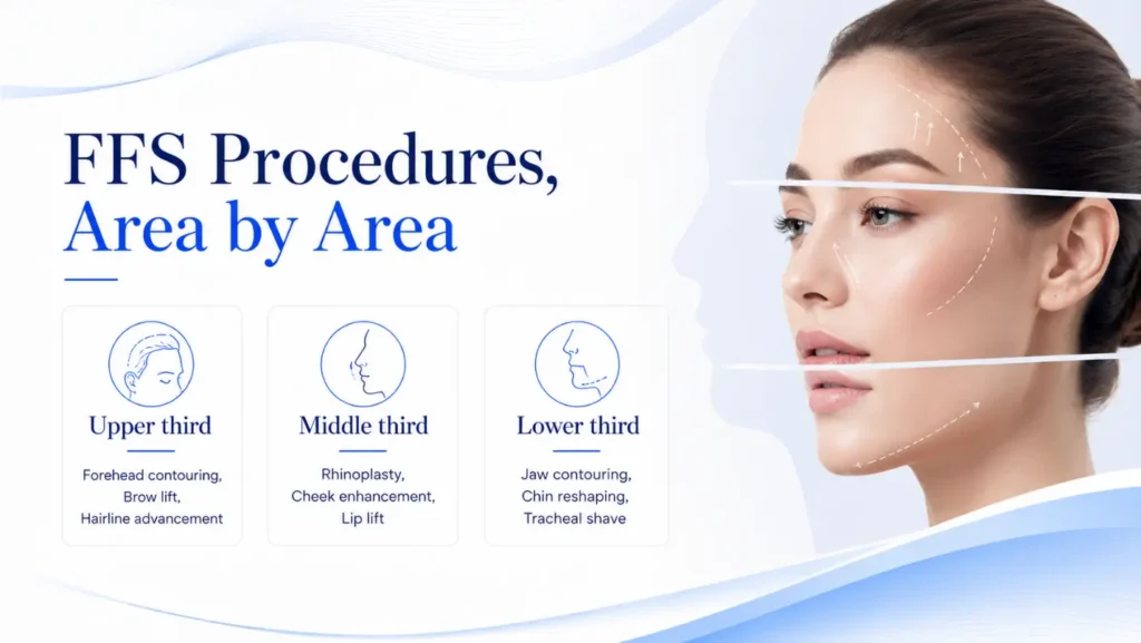
Before discussing what planning software measures, it is worth being clear about what it is measuring toward. Feminisation is often described as reduction, and that description quietly misleads. Some features are reduced — brow bossing, jaw angle width, chin height. Others are unchanged. Some are increased: cheek projection is often augmented, and the upper lip is frequently shortened to show more pink lip rather than made smaller.
What changes is the relationship between features and the proportion of the facial thirds. A forehead that has been set back alters how prominent the nose appears without the nose being touched. Reducing the height of the chin changes apparent facial length, which changes how the eyes read. This is why a plan built feature by feature produces a face that is smaller but not obviously more feminine, and why the measurements that matter are relational rather than absolute.
This is also the strongest argument for planning digitally. A 3D model allows those relationships to be measured and adjusted together before anyone operates, which is difficult to do reliably from photographs and clinical examination alone.
What the Accuracy Data Actually Shows
Here the evidence is more interesting than the marketing. A 2026 study from Yale published in Plastic and Reconstructive Surgery compared virtual surgical plans against post-operative CT imaging in 80 FFS patients, measuring how closely the actual result conformed to the plan. The answer differed sharply by procedure.
| Procedure | Volume conformity to plan | Interpretation |
| Forehead / frontal sinus setback | 92.9% | Highly predictable — the plan closely matches the result |
| Genioplasty (chin) | 81.8% | Good, but measurably less precise than forehead work |
| Gonial angle reduction (jaw) | 57.3% | Substantially less predictable — plan and result diverge |
The differences between forehead and the other two were statistically significant (p<0.0001), and the same pattern held for surface-area conformity: 95.8% for forehead, 89.6% for genioplasty, 73.0% for gonial angle. Planning accuracy is not one number. It depends on what is being planned.
| Why that pattern makes anatomical sense Forehead contouring involves a defined bony segment in an accessible, relatively simple geometry — the anterior wall of the frontal sinus is removed, reshaped and fixed. The plan translates cleanly. Gonial angle reduction is the opposite. The jaw angle is curved in three planes, access is limited, the bone is approached from inside the mouth, and the surgeon is often burring a contour rather than making a discrete cut. Reproducing a digital curve on real bone through a small intraoral opening is genuinely harder, and the data reflects that. A clinic that quotes a single accuracy figure across all FFS procedures is either unaware of this or not telling you. |
A 2025 systematic review conducted to PRISMA methodology, searching MEDLINE, EMBASE, Scopus, Web of Science and the Cochrane library, reached a broadly supportive but appropriately cautious conclusion: virtual surgical planning in osseous FFS was associated with reduced operative time, improved osteotomy accuracy and favourable patient-reported outcomes — while noting that “methodological heterogeneity and limited comparative data constrain definitive conclusions.” That sentence is worth reading twice. The evidence is encouraging and it is not yet conclusive.
Which Procedures Depend Most on Skeletal Planning
- Forehead contouring and frontal sinus setback. The strongest case. Imaging tells the surgeon how thick the anterior sinus wall is and how far the sinus extends — information that determines whether the bone can be set back at all, or only burred. This is a safety decision as much as an aesthetic one, and it cannot be judged from the outside of the head.
- Orbital rim contouring. Planned alongside the forehead; the rim and the brow ridge are a continuous surface and treating them as separate targets produces a visible step.
- Genioplasty. Planning defines how far the chin segment moves and in which planes. Related skeletal work is covered on our jaw and chin surgery page.
- Jaw angle reduction. Planning still helps — particularly for asymmetry and for mapping the inferior alveolar nerve — but the accuracy data above should temper expectations about millimetre reproduction.
- Rhinoplasty. Least dependent on skeletal planning. The nose is largely cartilage and soft tissue, judged intraoperatively. See rhinoplasty for how that assessment works.
- Cheek augmentation. Where a patient-specific implant is used, planning is central. Where fat grafting is used, it is not.
What 3D Planning Cannot Do
This section matters more than the one above it.
- It cannot predict your soft tissue precisely. Bone is planned; skin, fat and muscle then settle over it. A study of soft-tissue prediction in orthognathic planning found correct prediction ranging from 69.4% to 96.0% across the face, and — revealingly — the upper face (cheek, nose, upper lip) was predicted more accurately than the lower face (lower cheek, lower lip, chin). The soft tissue that moves most is predicted least well.
- It cannot guarantee symmetry. Most faces are asymmetric to begin with. Planning can measure and partially correct that; it cannot make a naturally asymmetric face symmetrical, and attempting it often looks worse than leaving a small difference.
- It cannot prevent complications. Planning reduces intraoperative uncertainty. It does not alter healing, infection risk, scarring or nerve recovery.
- A simulation image is not a promise. If a clinic shows you a simulated ‘after’ face, treat it as a way of discussing goals, not as the result you are buying. Ask directly whether it is a soft-tissue simulation or an artistic rendering — the two are frequently conflated.
- It does not replace clinical judgement. Research quantifying FFS changes has noted that a single skeletal change can produce diverse effects on soft tissue and on multiple cephalometric measurements at once. Software models that; it does not decide what is desirable.
Does It Change the Result, or the Process?
An honest answer, which few pages give: for much of FFS, virtual planning changes the process more than it changes the ceiling of what a skilled surgeon can achieve. Experienced craniofacial surgeons produced good FFS results before this technology existed, and still do.
What planning reliably contributes is different: shorter operative time, fewer intraoperative decisions taken under pressure, better preparation for anatomical surprises, more reproducible outcomes between cases, and a clearer conversation with the patient beforehand. In asymmetric and revision cases — where the anatomy is unusual and intraoperative judgement is hardest — the contribution is larger.
That is a real benefit. It is simply not the same claim as “3D planning makes surgery more accurate,” and the distinction is worth preserving.
Questions Worth Asking Any FFS Surgeon About Planning
- Will my frontal sinus be imaged before you decide the forehead technique?
- Is the plan transferred into theatre — through cutting guides, patient-specific implants or navigation — or does it stay on the screen?
- If you show me a simulation, is that a soft-tissue prediction or an illustration of the goal?
- How closely have your recent results matched your plans, and does that differ between forehead and jaw work?
- What will you do if the anatomy in theatre differs from the scan?
Frequently Asked Questions
What is virtual surgical planning in Facial Feminization Surgery?
It is the use of a three-dimensional scan of the facial skeleton to plan the operation before surgery — measuring bone thickness and position, deciding where cuts will be made and how far segments will move. It is distinct from 3D photography, which records the surface, and from simulation software, which suggests how a face might look afterwards.
Does 3D planning improve the accuracy of FFS?
Published evidence suggests it improves osteotomy accuracy and reduces operative time, though comparative data remains limited. Accuracy also varies considerably by procedure: one study of 80 patients found forehead volume conformity to plan of 92.9%, compared with 81.8% for genioplasty and 57.3% for gonial angle reduction.
Can 3D simulation predict exactly how I will look?
No. Bone position can be planned precisely; the soft tissue that drapes over it cannot. Soft-tissue prediction studies report accuracy ranging from roughly 69% to 96% across different facial regions, with the lower face predicted least reliably. A simulation is a tool for discussing goals, not a preview of your result.
Which FFS procedures benefit most from 3D planning?
Forehead contouring and frontal sinus setback benefit most, because imaging determines whether the anterior sinus wall can safely be set back and how far. Orbital rim work and genioplasty follow. Rhinoplasty benefits least, since the nose is mainly cartilage and soft tissue judged during surgery.
Is 3D planning available for FFS in India?
Yes — CT and cone-beam imaging, digital planning and patient-specific implants are available in Indian craniofacial practice, though the extent varies by clinic. The useful question is not whether a clinic has the technology but how the plan reaches the operating room.
Does 3D planning make surgery safer?
It can reduce intraoperative uncertainty, particularly around the frontal sinus and the nerves supplying the lower lip, and shorter operative time carries its own benefit. It does not alter healing, infection risk or scarring, and it does not remove the need for surgical experience.
Do I need 3D planning to get a good FFS result?
Not necessarily. Skilled craniofacial surgeons produced good results before this technology existed. Planning contributes most in asymmetric cases, revision surgery and complex skeletal work. It is a tool that supports judgement rather than a substitute for it.


